Fig. 3.
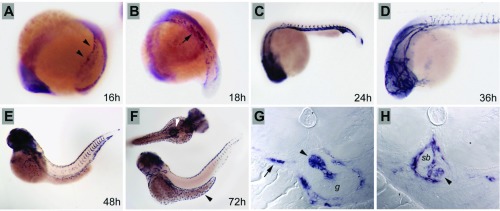
Expression of Aqp1a during embryogenesis. A: during somitogenesis, Aqp1a is expressed in the lateral mesoderm (arrowheads) and later is observed in cells converging at the midline to form the major trunk vessels (B; arrow). C: strong expression is observed at 24 hpf in the vasculature, including the forming intersomitic vessels. D: cranial vessels at 36 hpf are clearly delineated by Aqp1a expression. E: by 48 hpf, Aqp1a expression is downregulated in the vasculature but persists in single erythrocytes, here in the dorsal vasculature. F: ionocytes in the dermis over the yolk express aqp1a at 72 hpf (black arrowhead), while cells around the forming swim bladder also strongly express aqp1a (white arrowhead). G: histological sections of the forming swim bladder at 72 hpf show strong expression of aqp1a in the pneumatic duct (arrowhead) linking the gut (g) and the swim bladder and in erythrocytes in vessels (arrow). H: sections of the 72-hpf swim bladder (sb) show expression in the pneumatic duct where it joins the swim bladder (arrowhead) and in the vasculature surrounding the swim bladder.

