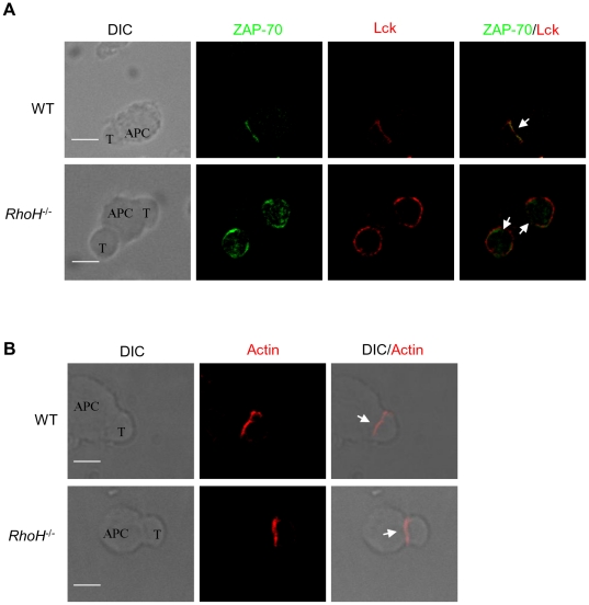
Figure 2. Impaired recruitment of Lck to the immunological synapse in Rhoh-/- T cells.
(A) CD8+ T cells from WT or Rhoh-/- p14 TCR transgenic mice were conjugated with gp33 peptide-preloaded APC cells (CH.B2 cells) for 5 min. Cells were fixed and stained with anti-ZAP-70 (green) and anti-Lck (red). The localization of the immune synapse is indicated with a white arrow. Bars, 3 µm. (B) T cell-APC conjugates were fixed and stained with TRITC-Phalloidin for detection of F-actin. Differential interference contrast (DIC) images show the antigen-specific T cell-APC conjugates. Bars, 3 µm. At least 100 cell conjugates were examined per condition.

