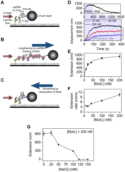
Figure 5. Single stranded DNA extension analysis of MutL binding activity.
(A) A 5.3 kb ssDNA is coiled randomly at a stretching force of 2.5 pN. (B) MutL binding extends the ssDNA and the position of the magnetic bead at the constant force of 2.5 pN. (C) Washing out free MutL results in gradual dissociation of the MutL bound to the ssDNA, resulting in the shortening of magnetic bead position. (D) Extension vs. time at different MutL concentrations (10, 40, and 200 nM). At 400 s, the free MutL was washed out of the flow chamber and the decrease in extension representing MutL dissociation monitored at 2.5 pN force. (E) Representative traces and the plot of extension (amplitude) measured at increasing concentrations of MutL. [MutL] = 10 nM (n = 51 beads), 20 nM (n = 15 beads), 40 nM (n = 52 beads), 100 nM (n = 37 beads), and 200 nM (n = 20 beads). (F) The extension rates measured at increasing concentrations of MutL. Studies in panels D–G were performed in the presence of 500 µM ATP in 25 mM NaCl. (G) The extension versus salt concentrations of 0 mM (n = 20 beads), 25 mM (n = 19 beads), 50 mM (n = 27 beads), 100 mM (n = 91 beads), 120 mM, and 150 mM NaCl in the presence of 200 nM MutL and 500 µM ATP. All the error bars represent s.e.m..

