Figure 3.
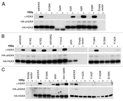
S139 phosphorylation and K119 monoubiquitination of H2AX mutants. H2AX−/− ES cells were transiently transfected with mammalian expression vectors encoding H2AX mutants shown and treated with IR as indicated 3 days after transfection. 30 minutes post-IR, histones were extracted for analysis by western blotting. γ-H2AX, HA-H2AX and HA-uH2AX are shown. Wild type H2AX (wtH2AX), S139A and empty vector were used as controls in each experiment. H2AX mutants DeIN (deletion of N-terminal 15 residues of H2AX), K5R, K9R and K36R are grouped in (A). K74Q, K75Q, KR75RQ (K75 to R and R76 to Q), T101A, 5KR (K118, 119, 127, 133 and 134 to R) and T136V as well as Y142A are grouped in (B). KK118RR (K118 and 119 to R), Y142F and Y142W as well as 5KR and Y142A are grouped in (C).

