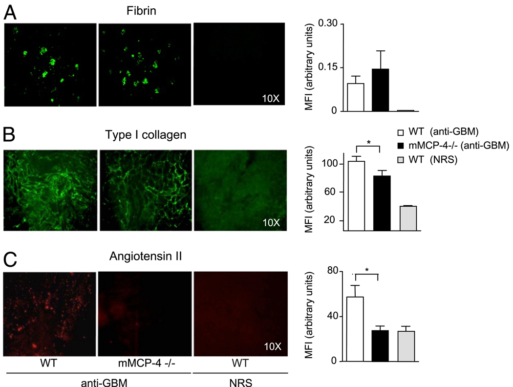
FIGURE 8.
mMCP-4–deficient mice have decreased glomerular and interstitial deposits of type I collagen and Ang II. At day 14, cryostat kidney sections from anti-GBM–stimulated mMCP-4−/− mice (n = 13) and WT mice (n = 9), as well as from NRS-treated WT control mice (n = 10) were stained with anti-fibrin Ab (A), anti-type I collagen Ab (B), or anti-Ang II Ab (C) and were analyzed by immunofluorescence light microscopy (left panels). Fluorescence intensities were quantified (right panels). Objective magnifications for glomerulus and interstitium are indicated in the right panels. Data are mean ± SEM per group. *p < 0.05.

