
Figure 4.
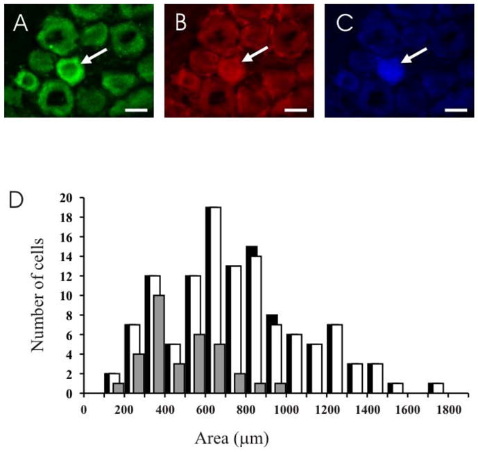
NMDA receptor expression in neurons that innervate the TMJ. The top panel consists of three images of the same field of a section through different filters to show a neuron (arrow) that is NR1 (A) and NR2B (B) immunoreactive, and contains Fast Blue (C). Scale bar = 20 μm. (D) Size distributions of neurons that innervate the TMJ (black), and TMJ afferents with NR1 (white) and NR2B (grey) subunit immunoreactivity.