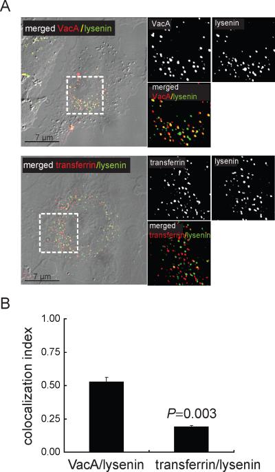
Fig. 1.
SM is associated with VacA-containing intracellular vesicles.
Co-localization of Alexa Fluor 647 labeled-VacA (50 nM) or Alexa Fluor 568 labeled-transferrin (60 nM) in AZ-521 cells with Venus-lysenin (1 μM) after 30 min was determined as described under Experimental Procedures.
A. Images were collected using DIC-fluorescence microscopy. The areas within the dashed white box were enlarged to illustrate VacA only (red puncta), transferrin only (red puncta), Venus-lysenin only (green puncta), colocalized VacA-Venus-lysenin (yellow puncta), or colocalized transferrin-Venus-lysenin (yellow puncta).
B. Co-localization analysis was carried out for the studies described in (A) as described under Experimental Procedures. The results shown were derived from data combined from three independent experiments. Statistical significance was calculated for differences in the colocalization index between those cells incubated with VacA and Venus-lysenin (VacA-Venus-lysenin) and those cells incubated with transferrin and Venus-lysenin (transferrin-Venus-lysenin).

