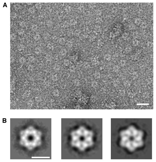
Figure 3. Two-dimensional structure of the yeast Rvb1/Rvb2 complex.
(A) Electron micrographs of negatively stained ring-shaped particles obtained upon incubation of the Rvb1 and Rvb2 proteins in the presence of ADP. Scale bar represents 200Å. (B) Two-dimensional averages of the yeast Rvb1/Rvb2 complex in the presence of ADP (left panel), ATP (center panel) and ATPγS (right panel). Scale bar represents 100Å.

