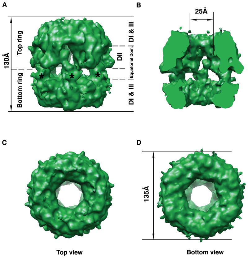
Figure 4. Cryo-electron microscopy 3D structure of the yeast Rvb1/Rvb2 complex.
(A) Side view of the three-dimensional structure of the Rvb1/Rvb2 complex. Top and bottom rings are indicated as well as the location of the DI, DII, DIII and equatorial domains of the monomers assembles within the dodecamer. The asterisks indicate the projected densities from the bottom ring into the equatorial domain. (B) The structure has been cut open to show the internal chamber, and the channel going through the structure. (C) Top view of the cryo-EM map. (D) Bottom view of the 3D EM map of the complex. The diameter of the structure is indicated.

