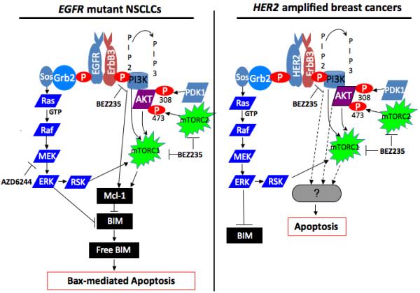
Figure 1. Differential survival signaling between EGFR mutant non-small cell lung cancers (NSCLCs) and HER2 amplified breast cancers.

(left diagram) In EGFR mutant NSCLCs, aberrant EGFR signal leads to activation of growth/survival pathways, most prominently PI3K and MEK/ERK signaling. PI3K signaling promotes Mcl-1 expression through a poorly defined mechanism that may be independent of AKT.6 Disruption of the signal by BEZ235 leads to loss of Mcl-1 expression, thereby increasing the levels of basal BIM not sequestered by Mcl-1. However, this effect in itself does not lead to sufficient unsequestered BIM to trigger substantial apoptosis. MEK signaling in these cells suppresses BIM expression by phosphorylation-mediated proteasome degradation.8 When this signal is interrupted by AZD6244, cellular BIM levels are increased. Again, this effect in itself does not lead to sufficient unsequestered BIM to promote apoptosis. However, simultaneous inhibiton of both pathways is necessary to yield sufficient unsequesterd BIM (“Free BIM”) to “prime” Bax,9 leading to mitochondrial-mediated apoptosis. (right diagram) In HER2 amplified breast cancers, aberrant HER2 signal leads to activation of growth/survival pathways, most prominently PI3K and MEK/ERK signaling. Disruption of PI3K/AKT/mTOR signaling alone is sufficient to induce substantial apoptosis. However, this is not associated with Mcl-1 loss. The mechanism leading to cell death remains poorly defined.
