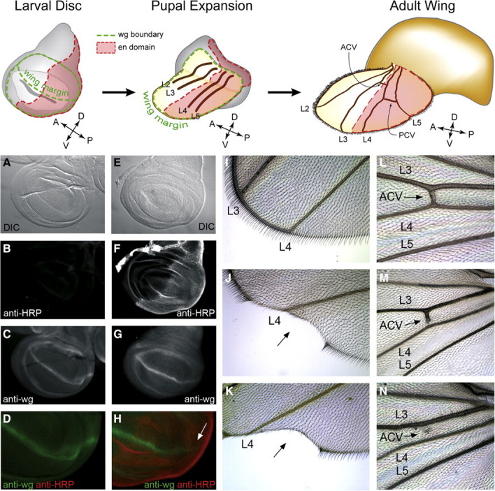
Fig. 8.

Ectopic α3-fucosylation in the posterior compartment of the wing disc alters Notch signaling and crossvein development. Reference figures across the top highlight the compartment boundaries that are established in the larval wing disc and illustrate their morphogenetic transformation into the adult wing. The posterior compartment of the wing disc is defined by the expression of engrailed (en, shown in red). In the adult wing, engrailed expressing cells contribute to the posterior half of the wing beginning at the intervein region between longitudinal veins L3 and L4. The anterior crossvein (ACV) derives from this region, while the posterior crossvein (PCV) arises from the intervein between L4 and L5. The dorsovental boundary in the larval wing disc prefigures the wing margin of the mature wing and is defined by expression of the wingless protein (wg, shown in green). Appropriate Notch signaling is essential for wg expression and the subsequent formation of the dorsoventral boundary. (A–D) Wild-type larval wing disc visualized by DIC (A), anti-HRP staining (B), anti-wg staining (C), and merge of anti-HRP/anti-wg (D). (E–H) Wing disc from en-GAL4; UAS-FucTA larva stained as in A–D. En-driven expression of FucTA attenuates wg expression along the dorsoventral boundary in the posterior compartment (arrow in H). (I–K) Appearance of the distal posterior wing margin in wild-type (I) and en-GAL4; UAS-FucTA (J, K) adults. With low penetrance (see Table I), the attenuation of wg expression observed in the larval wing disc results in wing notching (arrow in J, K). (L–N) The anterior crossvein (ACV) in wild-type wings interconnects L3 and L4 (L). In en-GAL4; UAS-FucTA wings, the ACV is frequently incomplete (M) and often completely missing (N).
