
Figure 2.
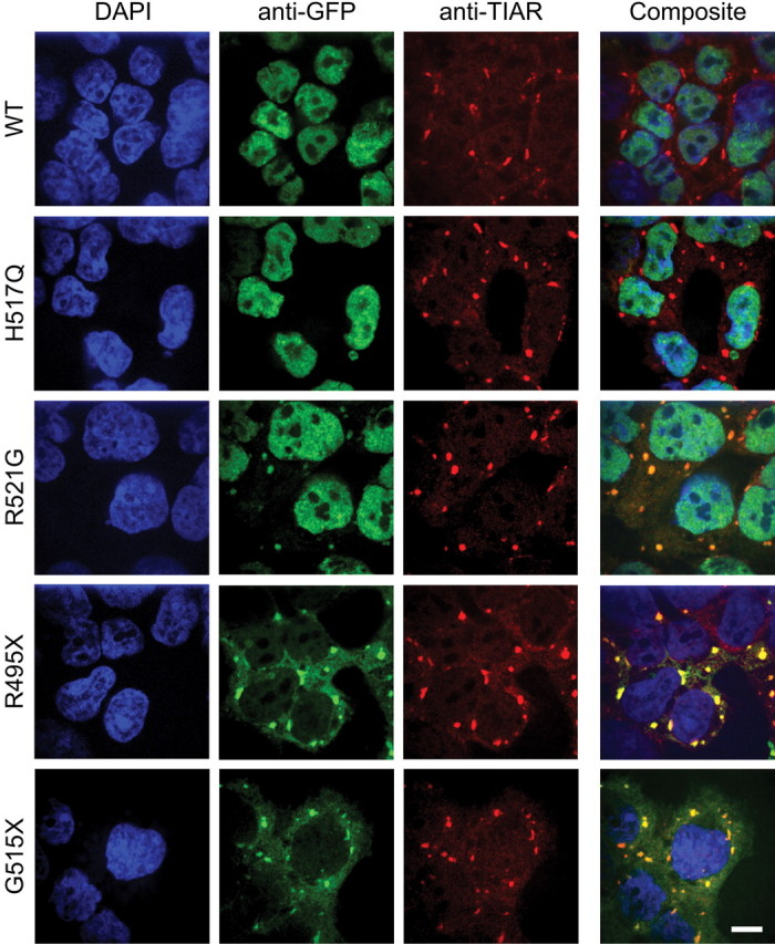
Incorporation of mutant FUS into stress granules in HEK-293 cells. Each GFP-FUS HEK-293 stable cell line was induced with doxycycline for 40 h, treated with 0.5 mm sodium arsenite for 1 h, and then probed with anti-GFP (green) and anti-TIAR (red) antibodies, and the nuclear dye DAPI (blue). Cytoplasmic aggregates containing GFP-FUS were detected with anti-GFP for the R521G, R495X and G5151X lines, but not for the WT and H517Q lines. Composite images indicate that the accumulated GFP-FUS(R521G, R495X and G515X) co-localized with the TIAR stress granule marker. Scale bar, 10 µm.