
Figure 7.
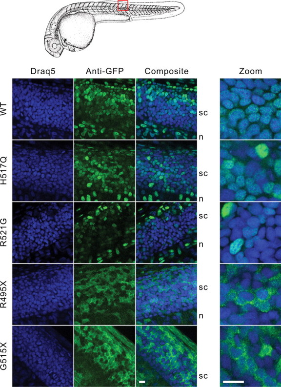
Expression of GFP-FUS variants in spinal cord of zebrafish embryos. Zebrafish eggs were injected with mRNAs encoding GFP-FUS variants, and embryos were processed for confocal microscopy (100×) as described in Figure 6. Shown are representative 0.9 µm slices (left panels) and 0.4 µm slices acquired using a 3.44× optical zoom (right panel). The higher magnification clearly showed the nuclear expression of GFP-FUS(WT, H517Q and R521G) variants and cytoplasmic accumulation of the R495X or G515X truncation mutants in the spinal cord. Scale bars, 10 µm.