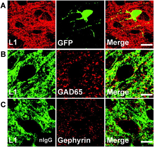
Figure 4.

L1 localizes to GABAergic inhibitory synaptic puncta in postnatal mouse cingulate cortex. (A) Double immunofluorescence staining of L1 and GFP in cingulate cortex (layers II/III) of WT/GAD67-EGFP mice at P10 showed L1 localized along processes and somal membranes of EGFP-positive basket interneurons. Scale bar: 20 μm. (B) Double staining of L1 and GAD65, a presynaptic inhibitory synapse marker, in cingulate cortex (layers II/III) of WT mice at P10 revealed L1 colocalized in part with GAD65. Scale bar: 10 μm. (C) Double staining of L1 and gephyrin, a postsynaptic inhibitory synapse marker, in cingulate cortex (layers II/III) of WT mice at P10 showed less colocalization of L1 with gephyrin. Scale bar: 10 μm.
