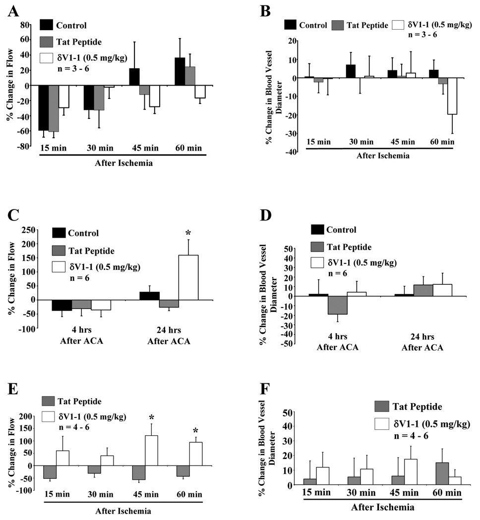
Figure 2.
δV1-1-induced attenuation of hypoperfusion 24 hrs after ACA. Similar experimental profile was used as described in Figure 1. 2-PM was used to image rat cortical microvessels before and after 6 min of ACA pretreated with tat peptide or δV1-1. Rats pretreated with tat peptide or δV1-1 was subjected to 6 min of ACA. 2-PM imaging analysis of cortical microvessels was performed before and after ACA (15 – 60 min) resulting in no significant change in overall cortical blood flow (A) or blood vessel diameter (B) (n = 3 – 6). Rats pretreated (30 min) with δV1-1 after 24 hrs of ACA significantly enhanced cortical microvessel blood flow by 160 % (C) with no change in blood vessel diameters detected 4 or 24 hrs after ACA in the presence or absence of δV1-1 (D) (n = 6). In a separate set of experiments, δV1-1 was administered directly after 6 min of ACA and CBF changes were monitored. CBF was enhanced 45 and 60 min after ACA (E), but no significant changes in blood vessel diameters were detected (n = 4 – 6) (F). * indicates p ≤ 0.05 as compared to tat peptide at the respective time point.

