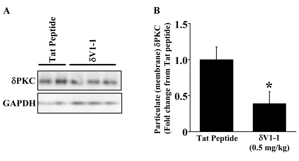
Figure 3.
δV1-1 inhibits δPKC translocation in cortical lysates. 24 hours after ACA, the animals were sacrificed and the cortex (site of 2-PM observed CBF) was extracted for Western blot analyses. Pretreatment with δV1-1 in the rat inhibited δPKC translocation after 6 min of ACA (A) with a 61 % reduction as compared to tat peptide (vehicle) treatment summarized in (B) (n = 4 – 6). Glyceraldehyde 3-phosphate dehydrogenase (GAPDH) was used as a loading control in all samples. * indicates p ≤ 0.05 as compared to tat peptide.

