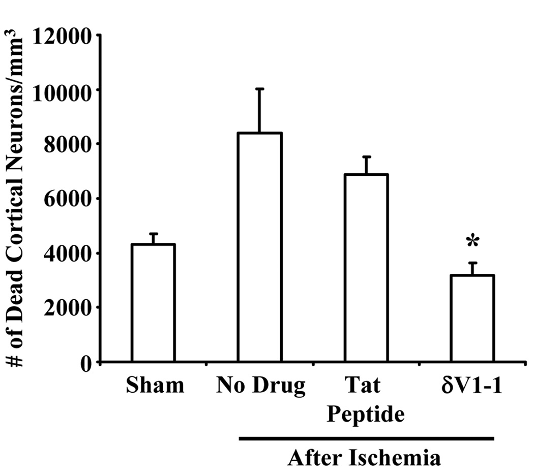
Figure 4.
δV1-1 provides neuroprotection in the cortex regions 7 days after ACA. Rats pretreated with δV1-1 or tat peptide were subjected to 6 min of ACA. Since cortical CBF was measured throughout this study (via 2-PM method), we determined the number of dead cortical neurons in the same region as CBF measurements. Rats pretreated with δV1-1 presented with lower number of dead cortical neurons as compared to no drug and tat peptide experimental groups after ACA (n = 3 – 11, * p ≤ 0.05).

