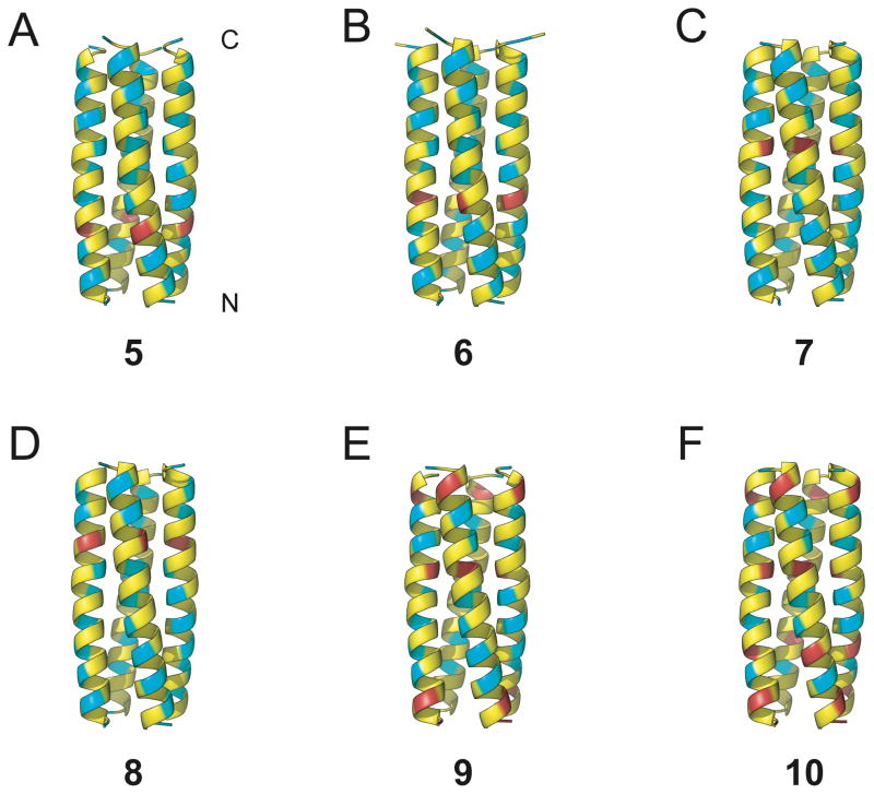
Figure 2.
Ribbon diagrams from x-ray crystal structures of α/β-peptides (A) 5 (PDB: 3HET), (B) 6 (PDB: 3HEU), (C) 7 (PDB: 3HEV), (D) 8 (PDB: 3HEW), (E) 9 (PDB: 3HEX), (F) 10 (PDB: HEY), showing the helix-bundle tetramer quaternary structures formed in each case. α-Amino acid residues are colored yellow, β3-amino acid residues are colored blue, and cyclic β-amino acid residues are colored red. Unlike 2, α/β-peptides 5–10 have a continuous heptad repeat pattern, without a helical stammer.

