
FIG. 6.
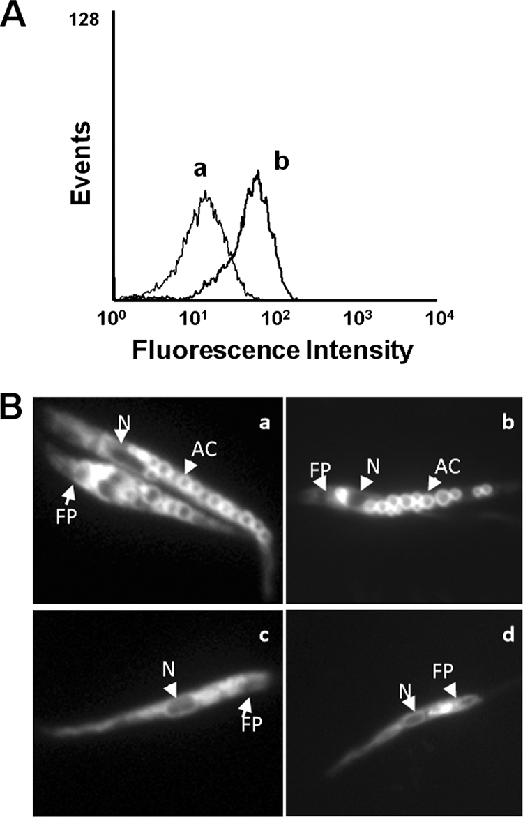
Intracellular localization of TFQ in Leishmania. (A) L. donovani promastigotes were pretreated with (a) and without (b) 5 μM TFQ for 10 min at 28°C and then labeled with 100 nM Lysotracker Green DND-26 for 10 min. Fluorescence was analyzed by flow cytometry as described in Materials and Methods. Representative histograms of three independent experiments are shown. (B) Intracellular localization of TFQ and Lysotracker Green in the control (a and b, respectively) and ΔAP3 (c and d, respectively) L. major promastigote lines were visualized by fluorescence microscopy after incubation with 5 μM TFQ or 100 nM Lysotracker Green DND-26 for 10 min at 28°C, as described in Materials and Methods. AC, acidocalcisome; N, nucleus; FP, flagellar pocket.