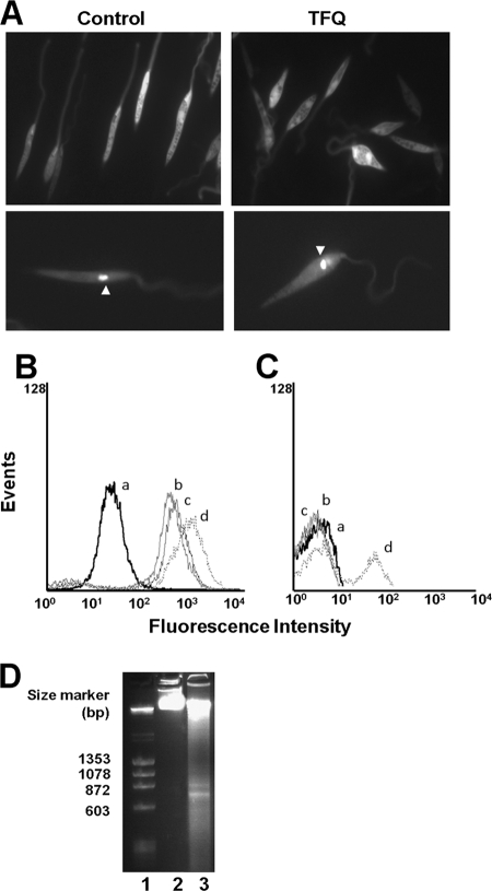
FIG. 7.
TFQ induces programmed cell death in Leishmania. (A) Autophagosome formation in L. donovani promastigotes. (Upper panel) Distribution of GFP-ATG8 in L. donovani promastigotes untreated or treated with 5 μM TFQ. (Lower panel) Autophagosomes could be identified as punctate structures clearly observable in the cytoplasm (arrowhead). (B and C) Representative histogram of TUNEL analysis and PI labeling, respectively, of L. donovani promastigotes treated with TFQ. Parasites were treated with different concentrations of TFQ: 5 (b), 10 (c) and 20 (d) μM for 4 h at 28°C, using untreated parasites (a) as controls. Fluorescein-dUTP and PI nucleic acid labeling were analyzed by flow cytometry as described in Materials and Methods. PI was used as the control of necrosis. Histograms are representative of three independent experiments with 10,000 parasites analyzed per group. (D) DNA fragmentation in L. donovani promastigotes. Genomic DNAs were isolated from parasites either untreated (lane 2) or treated for 4 h with 5 μM TFQ in HBS (lane 3), run through a 2% agarose gel, and visualized by ethidium bromide as described in Materials and Methods. DNA size markers (lane 1) are shown in base pairs (bp).

