
FIG. 6.
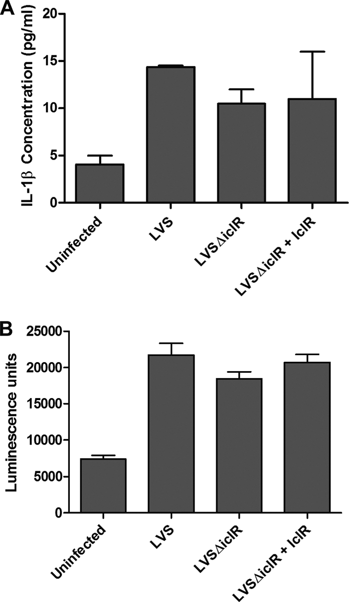
IL-1β release and cytotoxicity in murine bone marrow-derived macrophages infected with LVS ΔiclR. Infections were carried out at an MOI of 500 for wild-type LVS, LVS ΔiclR, and LVSΔiclR plus IclR (complementation). (A) IL-1β was quantified via ELISA, and (B) cytotoxicity was quantified via the ToxiLight bioassay (Lonza), both at 24 h postinfection. Graphs are representative of at least three separate experiments, with duplicate or triplicate wells for each strain per experiment. No differences were significant by any strain comparison based on Student's t test.