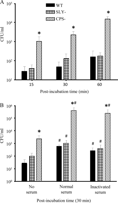
FIG. 1.
Phagocytosis of S. suis by murine microglial cells. (A) Kinetics of phagocytosis of S. suis strains (1 × 10⁶) by murine microglia after 15-, 30-, and 60-min infection times. *, P < 0.05 compared to phagocytosis levels obtained with the wild-type strain. (B) Effect of opsonization on phagocytosis at 30 min postinfection. Bacteria were nonopsonized (no serum) or preopsonized with 20% either normal or inactivated mouse serum. *, P < 0.05 compared to phagocytosis levels obtained with the wild-type strain; #, P < 0.05, indicating statistically significant differences between nonopsonized strains and their respective normal-serum- or inactivated-serum-opsonized counterparts. The numbers of internalized bacteria were determined by quantitative plating after 1 h of antibiotic treatment, and the results are expressed as CFU of recovered bacteria per ml (means plus SEM obtained from three independent experiments).

