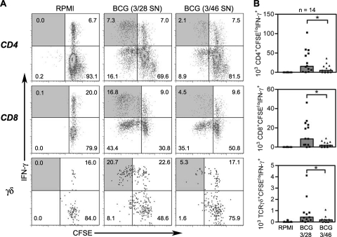
FIG. 2.
Flow cytometry to detect antimycobacterial CD4+, CD8+, and γδ TCR+ T cells and suppression by 3/46 supernatants. (A) CFSE-labeled PBMC were cultured in RPMI complete medium (left), in control 3/28 SN cells infected with BCG (middle), or in cells from 3/46 SN and infected with BCG (right). (B) Responding CD3+ CD4+, CD3+ CD8+, or CD3+ γδ+ T cells were enumerated by determining absolute numbers of CFSElo IFN-γ+ T cells after 7 days in culture. Bar graphs indicate the medians of 14 experiments. Asterisks indicate significant differences from T cell responses in 3/28 SN (P < 0.002 for all T cell subsets by Wilcoxon matched pairs test).

