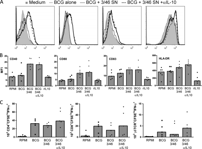
FIG. 7.
Dendritic cell maturation and antigen-presenting function are unaffected by 3/46 supernatants. DC were preincubated overnight in RPMI medium alone (no BCG), RPMI with BCG, 3/46 SN with BCG with or without neutralizing anti-IL-10, or RPMI with recombinant human IL-10 (10 ng/ml). DC were then analyzed by flow cytometry to measure cell surface expression of CD40, CD80, CD83, and HLA-DR as indicators of maturation. 3/46 SN did not prevent maturation of DC, as might be expected due to its high IL-10 content; in fact, the 3/46 SN increased expression of maturation markers. (A) Representative histograms indicate surface staining of DC incubated in medium alone (shaded histogram), with BCG (dotted line), BCG and 3/46 SN (solid line), or BCG plus 3/46 SN plus anti-IL-10 antibody (dashed line). (B) Mean fluorescence intensity (MFI) values for six individuals, with medians indicated by bars. (C) DC were infected with BCG overnight in the presence of RPMI or 3/46 SN with or without neutralizing anti-IL-10 antibody and then were washed to remove extracellular BCG. Autologous CFSE-labeled PBMC were then added to detect DC antigen presentation to mycobacterium-specific T cells. After 7 days, CD4+, CD8+, and γδ T cells responding to BCG (CFSElo IFN-γ+) were detected in similar absolute numbers whether or not DC were pretreated with 3/46 SN. Bars indicate medians for the data generated from six individuals.

