FIG. 1.
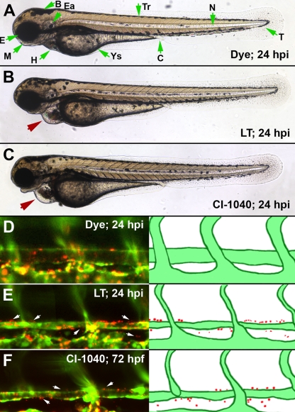
MEK1/2 inhibition phenocopies LT vascular permeability. An inhibitor for MEK1/2 produces a faithful phenocopy of LT vascular effects. CI-1040 was added to the medium of embryos at the same developmental time point at which we inject LT (48 hpf) and was washed out 6 h later. At 72 hpf, the embryos had pericardial edema, enlarged heart chambers, and vessel collapse (C), all phenotypes that are present in LT-injected embryos (B). In addition, both LT-injected and CI-1040-treated embryos had increased vascular permeability, as detected by the extravasation of 500-nm-diameter microspheres at 72 hpf (E and F). An LT dose of 150 fmol LF and 100 fmol PA was used in these experiments. Red arrows point out the pericardial edema noted in LT- or CI-1040-treated embryos, and white arrows mark regions of extravasated beads. Green arrows indicate morphological features of the zebrafish embryo (A). Abbreviations: B, brain; C, cloaca; Ea, ear; E, eye; H, heart; M, mouth; N, notochord; T, tail; Tr, trunk; Ys, yolk sac.

