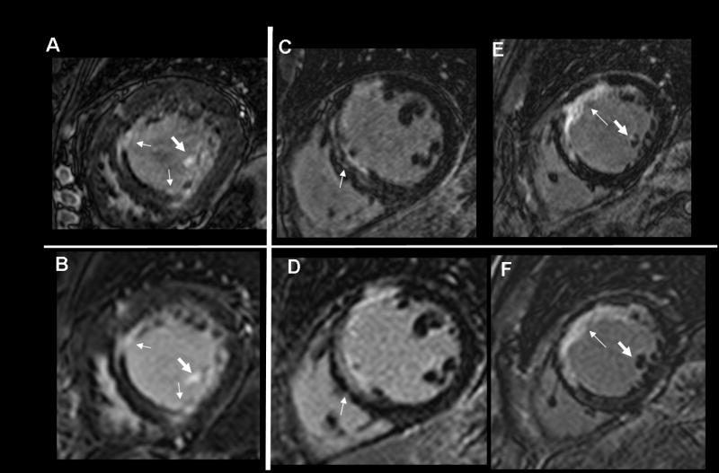
Figure 2.
Comparisons of 3D (A, C, E) and 2D (B, D, F) LGE in three subjects, showing higher spatial resolution provided by 3D LGE, illuminating an additional layer of complexity of myocardial scars (thin arrows). Subject 2 shows a double rim of scar (C, arrow), only suggested in (D). Papillary muscle LGE conspicuity is improved (fat arrows, subjects 1). The 2D images of subject 3 (F) do not demonstrate papillary muscle LGE, but some LGE is observed in an adjacent 2D slice.

