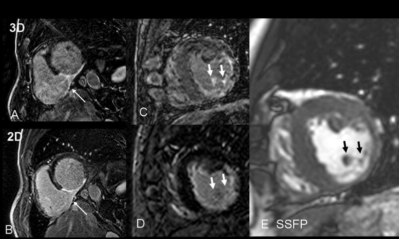
Figure 3.
A) Example of low quality 3D, here caused by excessive motion artifacts, compared to the good quality 2D LGE scan (B). Both images provide similar diagnostic information on inferior myocardial scar. The rare visualization of right ventricular infarct (arrow) is better observed in the lower quality 3D scan, likely due to the thinner slices and longer delay post injection. This patient has a homogeneous LGE structure. C) 3D LGE with imperfect myocardial nulling. Papillary LGE (arrows) was observed in the 3D but not the 2D scan (D), in a patient with a heterogeneous LGE structure. Although no non-invasive gold standard exists for detection of papillary muscle scar, the presence of LGE in many slices of the 3D volume at the anatomic site of the papillary muscle (E, arrows) is compelling.

