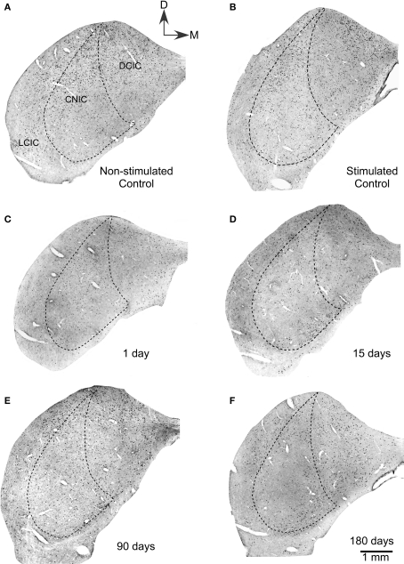
Figure 1.
Low power magnification photomicrograph showing sections of the ipsilateral inferior colliculus immunostained for c-Fos after lesions of the auditory cortex. (A) Non-stimulated control groups showed neuronal nuclei c-Fos Ir in all subdivisions of the IC. Also, in stimulated controls (B), a large number of c-Fos immunoreactive nuclei are seen in all three subdivisions of control inferior colliculi after sound stimulation. (C) One day after the cortical lesion, sound stimulation induces lower numbers of c-Fos immunoreactive neuronal nuclei. (D) Fifteen days after cortical ablation, sound-evoked c-Fos immunoreactivity is even lower than at 1 day after the lesion. (E) Ninety days after the cortical lesion, sound stimulation induces c-Fos immunoreactivity in neuronal nuclei, with numbers higher than at 15 days, but still lower than in stimulated controls. (F) Hundred eighty days after the cortical ablation, sound-evoked c-Fos immunoreactivity is close to control values.

