Figure 4.
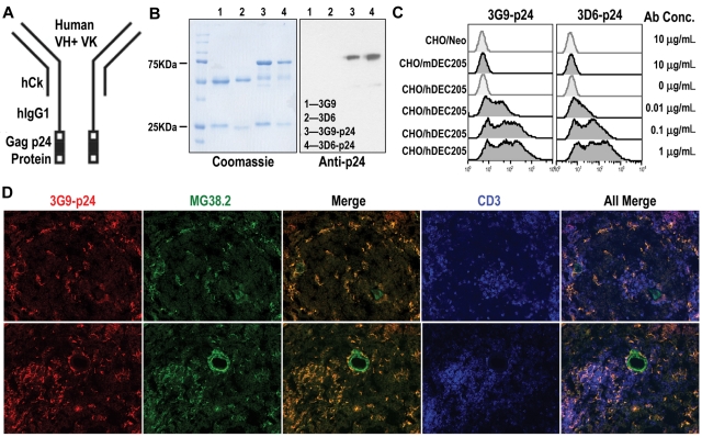
Expression and binding of hybrid anti-hDEC205-HIV Gag p24 fusion mAbs. (A) Schematic view of engineered mAb fused with HIV Gag p24. (B) Purified anti-hDEC205 mAbs 3D6 and 3G9 and their p24-fused hybrid mAbs were separated on 10% SDS-PAGE and stained with Coomassie blue (left) and blotted with anti-HIV Gag p24 antibodies (right). (C) Binding of hybrid mAbs to CHO/hDEC205, CHO/mDEC205, and control CHO/Neo cells using graded doses of purified recombinant p24-fused mAbs, followed by anti-hIgG-PE. (D) Detection of hDEC205 in normal human spleen sections after costaining with biotinylated 3G9-p24 (Alexa 555, red), MG38.2 mouse anti-hDEC205 (Alexa 488, green), and anti-hCD3 (APC, blue) to mark the T-cell areas. Images were taken 200× magnification using a Molecular Devices OlympusAX70 deconvolution microscope running METAMORPH Meta Imaging 3.0

