Figure 6.
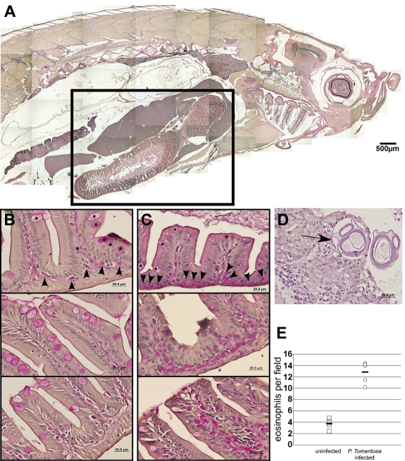
In vivo infection by P tomentosa leads to colonization of the gut and peritoneal cavity and an increase in local eosinophil number. (A) Composite image of sagittal section of adult zebrafish, stained with PAS. Inset: Area including intestines, shown in close-up views in panels B and C. (B) Imaging of the gut in uninfected animals shows nucleated PAS+ eosinophils interspersed along the basal lamina propria (arrowheads). Intestinal goblet cells (asterisks) are also PAS+, but distinct from eosinophils. (C) Infected animals show an increased number of PAS+ eosinophils along the basal lamina propria and also distally localized into villi. (D) P tomentosa larvae are observed within gut tissue (arrow). (E) Counts of eosinophils increase approximately 3-fold in intestines of infected animals. Images in panels A through D were acquired with an Olympus DP70 microscope and collected with Olympus DP Controller Version 2.1.1.183 and processed with Adobe Photoshop CS. Panel A is a composite image of a PAS and hematoxylin-stained section taken with a 10× dry objective. Images in panels B through D were taken with a 100× oil immersion objective and stained with PAS and hematoxylin.

