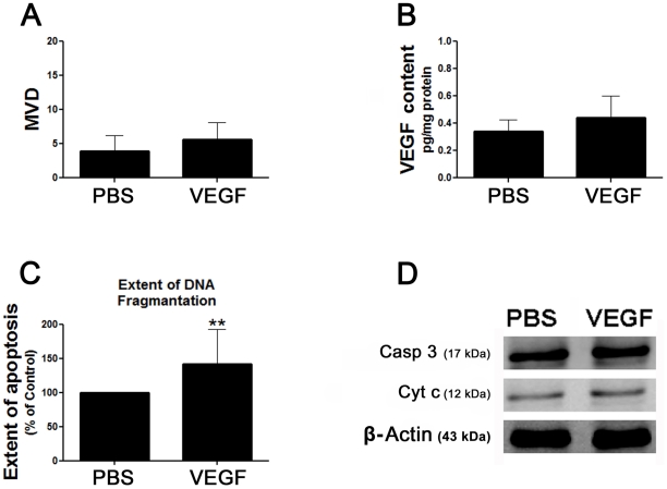
Figure 5. Effect of VEGF on MVD and the extent of apoptosis in the fat grafts.
PBS (100 µl) or VEGF (200 ng VEGF/100 µl PBS) were injected into the fat grafts in two different groups of mice on the day of the fat injection and every three days for 18 days. (A) Each bar represents the mean MVD ± SD from five regions of interest in each slide that was prepared from the harvested fat grafts of each treatment group at the end of the 15-week study period. (B) Each bar represents the mean VEGF content ± SD in the harvested fat grafts in each treatment group at the end of the 15-week study period. (C) The extent of apoptosis was measured by the TUNEL assay, and is expressed as a percentage of the extent of apoptosis in the PBS-treated fat grafts. Each bar represents the mean extent of apoptosis ± SD in the fat graft in each treatment group at the end of the 15-week study period. **P<0.01for the difference between the VEGF-treated fat grafts and the PBS-treated grafts. (D) Representative western blots of the expression levels of caspase 3 (Casp 3) and cytochrome c (Cyt c) in the PBS- and VEGF-treated fat grafts at the end of the 15-week study period.

