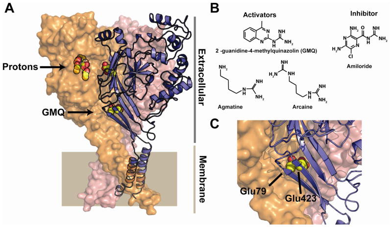
Figure 1.
A, Structure of ASIC1 (3HGC) (Gonzales et al., 2009). Conserved acidic pairs in the putative proton sensing pocket and those that affect GMQ action are shown in yellow and red. Putative proton sensing pocket and the site of GMQ covalent modification are shown. Extracellular and membrane-spanning regions are indicated. B, Chemical structures of GMQ, agmatine, arcaine, and amiloride. C, Closeup of the positions of the conserved Glu79 and Glu423 residues. The structure is from ASIC1. The labels correspond to the ASIC3 numbering.

