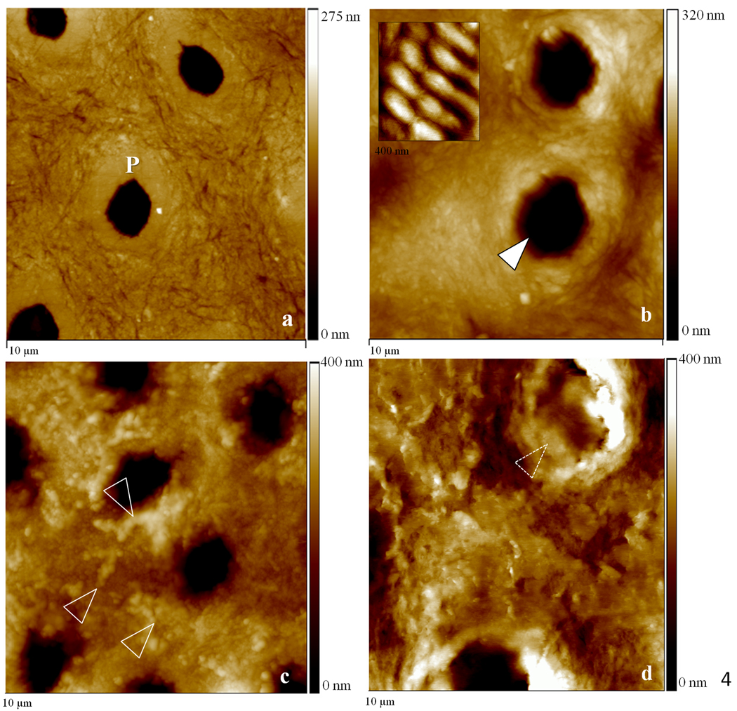
Figure 4.
10 µm AFM scans in the contact mode of specimens remineralized for five consecutive days as well as normal and demineralized specimens. (a) Normal dentin showing peritubular mineral surrounding tubules (P); (b) demineralized dentin exhibited surface roughened topography and widened tubular lumens (arrowhead). Fully demineralized collagen from dentin is shown in the detail for comparison purposes (400 nm scan); (c) remineralization with continuous approach suggested mineral in association with the demineralized organic network, white arrowheads show zones suggestive of mineral attached to the collagen network. (d) Remineralization using static approach showed random precipitation of mineral onto the surface of dentin with no preferential organization;

