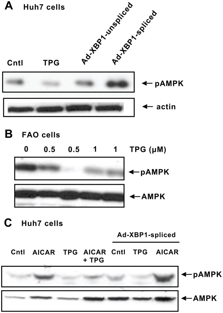
Fig. 1.
ER stress down-regulates pAMPK levels. A, Huh7 cells were treated with 300 nM TPG or infected with Ad-XBP1-unspliced or Ad-XBP1-spliced. Cytosolic proteins were used for Western blot analysis to detect pAMPK and β-actin levels. B, TPG (500 nm and 1μM) suppresses pAMPK levels in FAO cells. pAMPK and total AMPK levels were detected in cytosolic proteins of control and treated cells after 24 h treatment with TPG. C, Huh7 cells were treated with 500 μM AICAR or 300 nM TPG alone or in combination for 2 h. The cells were also infected with Ad-XBP1-spliced for 24 h followed by treatment with either TPG or AICAR for 2 h. Cytosolic proteins were run on SDS-PAGE to detect pAMPK and total AMPK levels. Blots shown are representative of three separate experiments with similar results.

