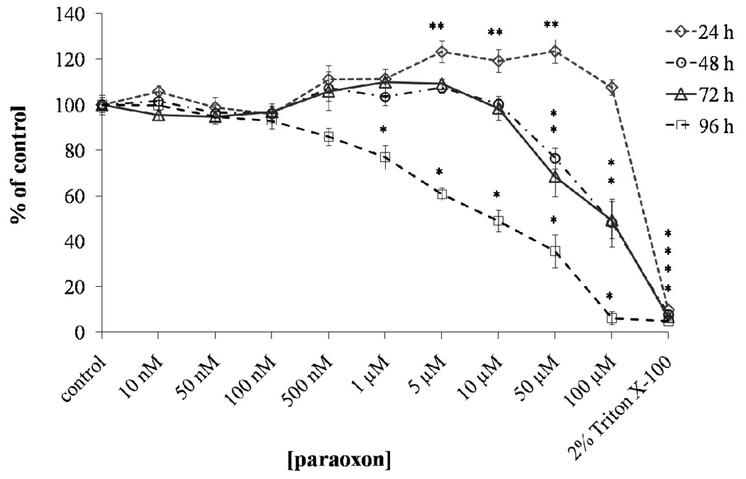
Figure 2.
Cytotoxicity of paraoxon to SH-SY5Y cells. Cells were treated with paraoxon (10 nM – 100µM) for 24, 48, 72 and 96 h or no paraoxon (control) and the cell viability estimated by 3-(4, 5-dimethylthiazol-2-yl)-2, 5-diphenyl tetrazolium bromide (MTT) colorimetric assay. Results are presented as % of vehicle control, determined by comparing the absorbance readings of the wells containing the paraoxon treated cells with those of the vehicle (0.1% acetonitrile) treated cells. Triton X-100 was used as a positive control (= 100% cell death). Significance determined by one-way ANOVA followed by Dunnet’s post hoc test (* significant decrease; ** significant increase) (p < 0.05; n = 8).

