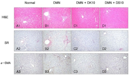
Fig. 1.
Histological analysis of liver sections. (A) Normal. (B) DMN (10 mg/kg per day for 3 consecutive days per week for 4 weeks) alone. (C) DMN with 10% grape skins. (D) DMN with 10% grape seeds. The sections were stained with hematoxylin-eosin (H&E) and with Sirius red (SR). Activated HSCs were detected by immunohistochemistry with α-SMA antibody (α-SMA).

