Figure 2.

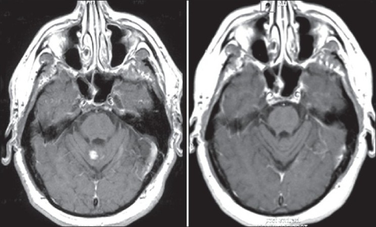
Axial T1-weighted postcontrast images demonstrating enhancing lesion in the vermis 8/06 (left) with resolution of the lesion post infl iximab 3/07 (right)

Axial T1-weighted postcontrast images demonstrating enhancing lesion in the vermis 8/06 (left) with resolution of the lesion post infl iximab 3/07 (right)