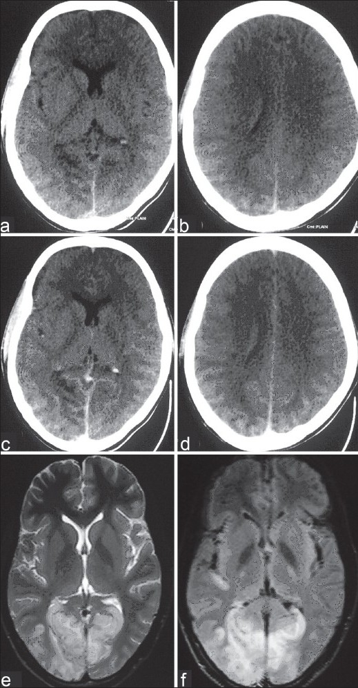
Figure 2.

(a–e) : CT scan (2nd December 2007) showing ill-defined bilateral right more than left occipitoparietal interdigitating hypodense lesions with effacement of adjacent sulci and no postcontrast enhancement. (e, f) MRI (29th November 2007) shows bilateral right more than left occipitoparietal T2W and FLAIR hyperintense lesions predominantly involving the white matter but also the gray matter in the right occipital area
