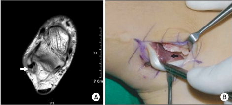
Fig. 1.
(A) A T2-weighted axial image of an ankle MRI showing crowding of the muscle tissue of the peroneus brevis tendon within the superior peroneal retinaculum (white arrow). (B) In the same area, extension of the muscle tissue of the peroneus brevis tendon distal to the fibular groove is observed (black arrow).

