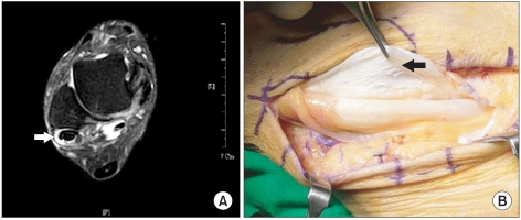
Fig. 4.
(A) This T2-weighted fat suppression axial image of an ankle MRI shows a split tear and enlarged shape of the peroneus brevis tendon (white arrow). Around peroneus tendons, an increase in synovial fluid is observed. (B) In the same area, a split tear of the peroneus brevis tendon and thickened synovium is observed (black arrow).

