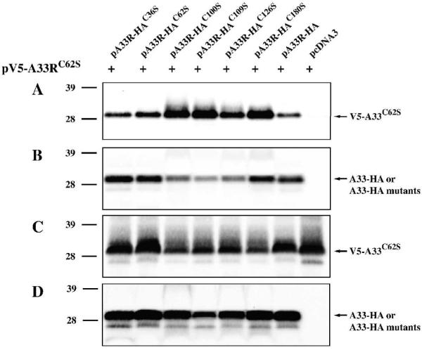
Fig. 3. A33-HAC62S forms dimers.
HeLa cells were infected with vTF7.3 in the presence of AraC and transfected with the indicated plasmids. 24 h PI, cells were harvested and lysed in RIPA buffer. A33-HA or A33-HA cysteine-to-serine mutants were immunoprecipitated with an anti-HA MAb. Immune complexes (A&B) or cell lysates (C&D) were resolved by SDS-PAGE. V5-A33C62S was detected by Western blotting with an HRP-conjugated anti-V5 antibody (A&C). After probing with an anti-V5 antibody, the blots were stripped and re-probed with an HRP-conjugated anti-HA antibody (B&D). The positions and molecular weights, in kDa, of marker proteins are shown.

