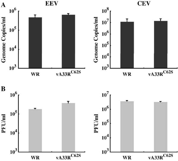
Fig. 8. vA33RC62S and WR produce similar amounts of EEV and CEV.
BS-C-1 cells were infected with either WR or vA33RC62S at a MOI of 10.0 in duplicate. 24 h PI, EEV containing supernatants were collected and cell monolayers were incubated with media containing 1 μg per ml of trypsin at 37°C for 1 h. Afterward, the media was collected (CEV). The amounts of EEV and CEV produced were measured by absolute quantification of genome copies using real-time PCR (A) and their infectivity was determined by plaque assay on fresh BS-C-1 cell monolayers (B). The averages of EEV or CEV and the error bars for each virus are plotted.

