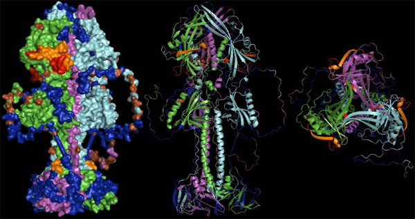
Figure 9.

Representation of the gp110 putative trimer surface and secondary structure cartoons in lateral and top view (left to right). Monomers are drawn in green, violet and cyan. Regions covered by predicted, potentially neutralizing epitiopes are shown in blue, residues predicted to be glycosylated are given in brown. Areas coded in red were experimentally shown to be neutralizing in homologous proteins of other herpesviruses, while areas coded in orange were additionally predicted as epitopes. The orange spot at the stem of the molecule indicates the terminus of a neutralizing epitope close to the N-terminus of the protein (unfortunately only partially resolved in the structure model).
