Figure 2.
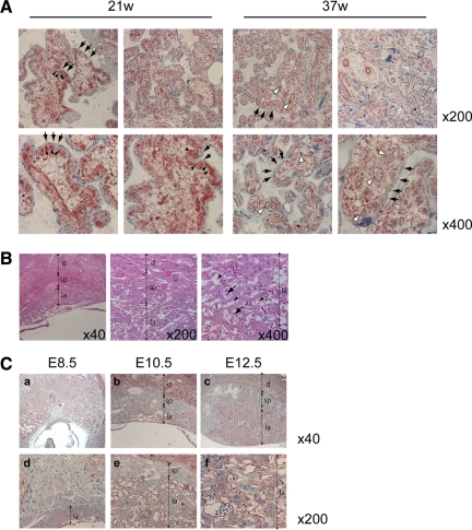
CNN3 expression in human and mouse placental tissues. (A) Immunohistochemistry of chorionic villi of human placentas at 21 and 37 wk of gestation. CNN3 was detected in the cytotrophoblasts (black arrowheads) and fetal endothelial cells (white arrowheads) but not in the syncytiotrophoblast layer (arrows). (B) Histology of mouse placenta. Transverse sections (4 μm) were stained with hematoxylin/eosin. A few maternal blood sinuses (arrowheads) and fetal blood vessels (arrows) are found within the labyrinth region. Localizations of the labyrinth layer (la), spongiotrophoblast layer (sp), and maternal decidua (d) are indicated. (C) Immunohistochemistry of mouse placental tissues at 8.5 dpc (a, d), 10.5 dpc (b, e), and 12.5 dpc (c, f). CNN3 was detected in the labyrinth layer and maternal deciduas.

