Figure 2.
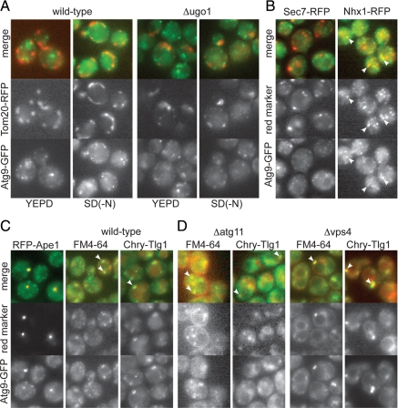
Intracellular distribution of Atg9-GFP. (A) Fluorescent micrographs comparing Atg9-GFP with the mitochondrial outer membrane marker Tom20-RFP in wild-type cells or cells lacking the mitochondrial fusion protein Ugo1 and grown in YEPD or starved for nitrogen for four hours [SD(-N)] as indicated. The loss of Ugo1 causes the mitochondria to form clumps of fragments (Sesaki and Jensen, 2001), but Atg9-GFP remains scattered, with colocalization only observed occasionally, usually in forming buds. (B) Fluorescence micrographs comparing Atg9-GFP to the late Golgi protein Sec7-RFP or the late endosomal protein Nhx1-RFP in wild-type cells in rich medium. (C) Fluorescence micrographs comparing Atg9-GFP to RFP-Ape1, the endocytic tracer FM4-64 (10 min pulse, 10 min chase), or mCherry-Tlg1 (Chry-Tlg1) in wild-type cells in rich medium. (D) As for C except that the cells are either lacking the autophagosome scaffold protein Atg11 which is required for membrane to accumulate at the autophagosome (Shintani and Klionsky, 2004), or lacking the endosomal protein Vps4, in which the endocytic compartment expands (Babst et al., 1997).

