Figure 6.
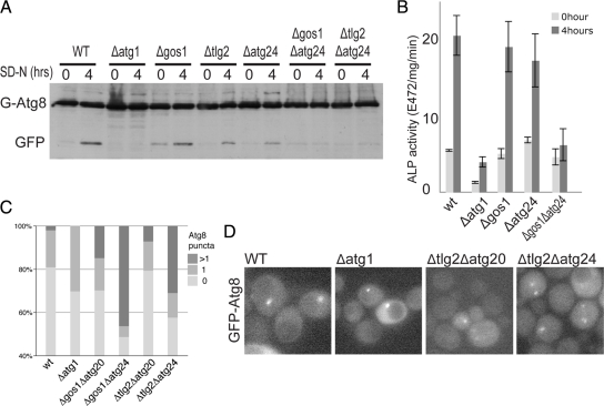
Combined mutants in Golgi-endosomal components show synergistic defects in starvation-induced autophagy. (A) Anti-GFP immunoblot of the indicated strains expressing GFP-Atg8. Cells were grown without uracil (rich medium) to midlog phase and either harvested or shifted to starvation conditions for four hours (SD-N). The positions of GFP-Atg8 and the free GFP that is released after autophagic delivery to the vacuole are indicated. (B) Alkaline phosphatase activity in the indicated strains expressing Pho8Δ60 that lacks a transmembrane domain and so is only activated after delivery to the vacuole by autophagy. Starvation and strains (with PHO13 deleted) are as in A, and error bars indicate the SD of three independent experiments. (C) Quantitation of the distribution of GFP-Atg8 in cells grown as in A. For each strain >150 cells were counted. The values show the proportion of the population with one, or more than one puncta of GFP-Atg8, and are based on a projections of six focal planes. The increases in frequency of cells with multiple GFP-Atg8 puncta in all the double mutants are statistically significant (χ2 test, two-tailed values p < 0.0001). (D) Representative single focal plane images of the cells quantified in C. Combination of SNARE and Atg24 mutations results in the appearance of multiple puncta of GFP-Atg8 consistent with defects in autophagosome formation.

