Figure 2.
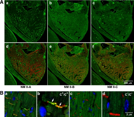
Expression of NM II-C in embryonic and adult mouse hearts. (A) Immunofluorescence confocal images of an E13.5 mouse heart stained for NMHC II-A (a, green), II-B (b, green), and II-C (c, green) together with desmin (d–f, red), a marker for (cardiac) myocytes. NM II-A is only expressed in nonmyocytes (a, green). Note the lack of colocalization with desmin-positive cardiac myocytes. NM II-B is detected in both myocytes (e, red and green colocalization) and nonmyocytes (e, green) in the heart. NM II-C is detected in myocytes (f, red and green colocalization) but not in nonmyocytes. The bright green spots are autofluorescence from red blood cells in c and f. (B) Immunofluorescence confocal images of adult heart sections from C+/C+ (a, magnified in b) and C−/C− (c, magnified in d) mice. N-Cadherin is a marker for the intercalated disc (red). Nuclei are stained with DAPI (blue). Arrows in b indicates the presence of NMHC II-C (green) in the intercalated disc. NMHC II-C is absent from C−/C− intercalated discs (c and d).

