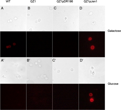
Figure 2.
Detection of JEN1 expression in yeast membrane by immunofluorescence. Immunofluorescence staining of the four yeast strains from Figure 1 was performed using cells grown in SD medium containing either 2% glucose or galactose, as indicated. Images of bright field and immunofluorescence fields are shown side by side.

