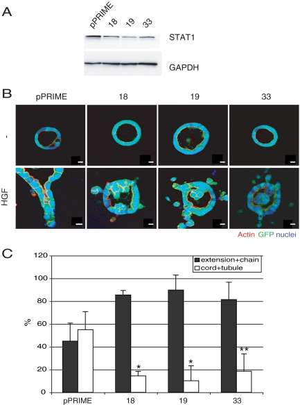
Figure 1.
Inhibition of STAT1 retards HGF-induced MDCK tubulogenesis. (A) Expression level of STAT1 in control (pPRIME) and STAT1 knockdown lines (18, 19, and 33) was compared by immunoblot with anti-STAT1 antibody. (B) Control cells or STAT1 knockdown cells were grown as cysts and either left untreated (top row) or treated with HGF for 72 h (bottom row). Inhibition of STAT1 blocks the HGF-induced redifferentiation phase. The GFP represents GFP-pPRIME transfectant. Bar, 10 μm. (C) Quantification of tubular structures in the presence of HGF. One hundred GFP-positive structures per sample were counted, and values are the mean ± SEM of three independent experiments. *p < 0.01 and **p < 0.05.

