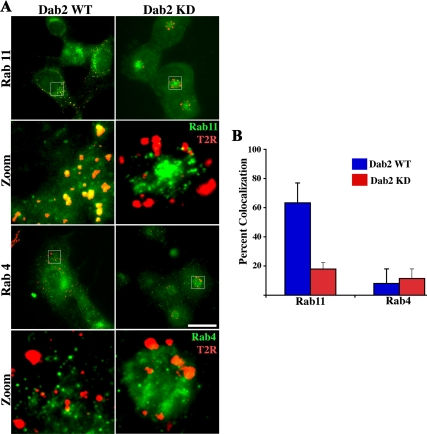
Figure 3.
TGF-β receptors localize to a Rab11-positive compartment in Dab2 WT cells but not in Dab2 KD cells. (A) Chimeric TGF-β receptors stably expressed in Dab2 WT (left) and Dab2 KD16 cells (right) were bound to anti-GM-CSFR-β antibody (recognizes the extracellular domain) at 10°C and internalized at 37°C for 45 min. Cells were then acid washed to remove remaining cell surface antibodies, fixed, permeabilized, and incubated with rabbit anti-Rab11 (top) and Rab4 antibody (bottom). The Rab11/Rab4 compartment was detected by incubation with Alexa Fluor 488-labeled (green) anti-rabbit, whereas internalized receptors (red) were visualized after treatment with Cy3-conjugated anti-mouse secondary antibodies, respectively. Separate images were acquired for each fluorophore via fluorescence microscopy. Images were collected in pseudocolor and are presented as overlays. (B) Percentage of colocalization of receptors with Rab11 and Rab4 and represents the mean ± SD for 50 cells from each of three independent experiments. Bar, 10 μm.

